Home » Uncategories » Abdominal Anatomy - Medically Accurate Illustration Of The Abdominal Anatomy Stock Photo Picture And Royalty Free Image Image 44543354
Thursday, April 22, 2021
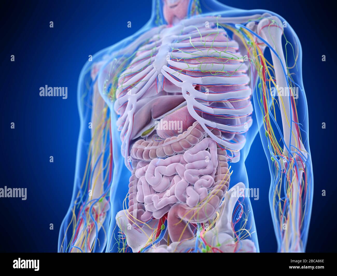
Abdominal Anatomy - Medically Accurate Illustration Of The Abdominal Anatomy Stock Photo Picture And Royalty Free Image Image 44543354
Abdominal Anatomy - Medically Accurate Illustration Of The Abdominal Anatomy Stock Photo Picture And Royalty Free Image Image 44543354. These organs are held together loosely by connecting tissues. It also contains the spleen. Abdominal anatomy includes a major element of the gastrointestinal, system, the caudal end of the oesophagus, stomach, large and small intestine, liver, pancreas and the gallbladder. Liver, spleen, diaphragm, stomach : The abdominal wall surrounds the abdominal cavity, providing it with flexible coverage and protecting the internal organs from damage.
ads/bitcoin1.txt
It is based on our previous model thorax and abdomen: These changes are subtle, but with practice, you should be able to make out several organs and muscles. Overview on what a radiologist is and what they do. Rectus abdominis, informally known as the abs muscle, is a long muscle of the anterior abdominal wall. This article is the detailed account of all the major organs that are categorized under the nine regions in the abdominal cavity 1) stomach 2) intestines a) small intestine duodenum jejunum ileum b) large intestine ceacum colon (ascending, transverse and descending) rectum anal canal 3) liver 4) gall bladder 5) pancreas 6) spleen 7) kidneys …
Anatomy Of Human Abdominal Vein System Wall Art Canvas Prints Framed Prints Wall Peels Great Big Canvas from static.greatbigcanvas.com This is a laparoscopic tour of abdominal cavity anatomy. The right upper quadrant primarily. In those with low body fat, it is clearly visible beneath the skin forming the 'six pack'. This article is the detailed account of all the major organs that are categorized under the nine regions in the abdominal cavity 1) stomach 2) intestines a) small intestine duodenum jejunum ileum b) large intestine ceacum colon (ascending, transverse and descending) rectum anal canal 3) liver 4) gall bladder 5) pancreas 6) spleen 7) kidneys … Now that we have divided the abdomen into different regions, let's discuss the anatomy of each section. The diaphragm is its upper boundary. Some arteries and veins. to better visualize the pancreas, some of the organs can be made transparent by scrubbing through the animation. 1 like other blood vessels, the wall of the abdominal aorta is made up of three distinct tissue layers:
We're going to take apart a plastic anatomy model and see what we can find in the abdomen.
ads/bitcoin2.txt
Liver, spleen, diaphragm, stomach : That have the ability to constrict and relax as needed to adjust for high and low blood pressures. We'll identify as many organs as we can, see how they fit into the. The coeliac trunk arises from the abdominal aorta at t12 and supplies the foregut gastrointestinal viscera. Setting up a radiology society. Arises anteriorly from abdominal aorta just below diaphragm at the t12 level, behind the median arcuate ligament, just as the aorta enters the abdomen in between right and left crura. The abdomen is the part of the body that contains all of the structures between the thorax (chest) and the pelvis, and is separated from the thorax via the diaphragm. Rectus abdominis, informally known as the abs muscle, is a long muscle of the anterior abdominal wall. The diaphragm is its upper boundary. This model shows some of the organs and vessels in the abdomen. This is a laparoscopic tour of abdominal cavity anatomy. Abdomen, in human anatomy, the body cavity lying between the chest or thorax above and the pelvis below and from the spine in the back to the wall of abdominal muscles in the front. Now that we have divided the abdomen into different regions, let's discuss the anatomy of each section.
A collection of articles covering abdominal anatomy, including abdominal wall anatomy and abdominal cavity anatomy. It also contains the spleen. The diaphragm is its upper boundary. The abdomen is the front part of the abdominal segment of the trunk. The abdomen contains all the digestive organs, including the stomach, small and large intestines, pancreas, liver, and gallbladder.
Male Abdominal Muscle Anatomy Muscle Anatomy Abdominal Muscles Anatomy Anatomy from i.pinimg.com Together, these three turn nutrients into usable energy, as well as help dispose of solid waste. That have the ability to constrict and relax as needed to adjust for high and low blood pressures. In those with low body fat, it is clearly visible beneath the skin forming the 'six pack'. Abdominal anatomy includes a major element of the gastrointestinal, system, the caudal end of the oesophagus, stomach, large and small intestine, liver, pancreas and the gallbladder. This is a laparoscopic tour of abdominal cavity anatomy. This article is the detailed account of all the major organs that are categorized under the nine regions in the abdominal cavity 1) stomach 2) intestines a) small intestine duodenum jejunum ileum b) large intestine ceacum colon (ascending, transverse and descending) rectum anal canal 3) liver 4) gall bladder 5) pancreas 6) spleen 7) kidneys … Rectus abdominis, informally known as the abs muscle, is a long muscle of the anterior abdominal wall. Injury to any region of the abdomen can of course create injury to any of the organs contained therein.
This is a laparoscopic tour of abdominal cavity anatomy.
ads/bitcoin2.txt
In those with low body fat, it is clearly visible beneath the skin forming the 'six pack'. Get the book for free. The major organs of the abdomen include the small intestine, large intestine, and stomach. This article is the detailed account of all the major organs that are categorized under the nine regions in the abdominal cavity 1) stomach 2) intestines a) small intestine duodenum jejunum ileum b) large intestine ceacum colon (ascending, transverse and descending) rectum anal canal 3) liver 4) gall bladder 5) pancreas 6) spleen 7) kidneys … The diaphragm is its upper boundary. The viewer gets to see the abdominal organs just as the surgeon does while he or she is operating o. By the time the aorta reaches the abdomen, it has tapered to a width of about 2 centimeters wide, making it the largest artery in the abdominal cavity. The abdominal portion of the aorta supplies most of the abdomen, and begins at the level of the twelfth thoracic vertebra (t12), and then terminates at l4 by bifurcating into the left and right common iliac arteries. Said to be present when the abdominal wall, having been compressed slowly, is released rapidly and results in sudden sharp abdominal pain. Liver, spleen, colon, bladder, stomach, pancreas. The abdominal cavity is the part of the body that houses the stomach, liver, pancreas, kidneys, gallbladder, spleen, and the large and small intestines.the diaphragm marks the top of the abdomen and the horizontal line at the level of the top of the pelvis marks the bottom. However, commonly injury to a specific area will result in injury to those organs contained within that locality. Setting up a radiology society.
The major organs of the abdomen include the small intestine, large intestine, and stomach. Also note that portions of structures can extend into other regions as well. This model shows some of the organs and vessels in the abdomen. The thin inner layer (tunica intima), the thick middle layer (tunica media), and the thin outer layer (tunica adventitia). The abdomen is the front part of the abdominal segment of the trunk.
Abdominal Anatomy Illustration Stock Photo Alamy from c8.alamy.com Together, these three turn nutrients into usable energy, as well as help dispose of solid waste. Overview on what a radiologist is and what they do. Get the book for free. Said to be present when the abdominal wall, having been compressed slowly, is released rapidly and results in sudden sharp abdominal pain. The below image can be used as a reference. The right upper quadrant primarily. The abdomen is the front part of the abdominal segment of the trunk. It is based on our previous model thorax and abdomen:
Liver, spleen, colon, bladder, stomach, pancreas.
ads/bitcoin2.txt
The abdominal cavity is the part of the body that houses the stomach, liver, pancreas, kidneys, gallbladder, spleen, and the large and small intestines.the diaphragm marks the top of the abdomen and the horizontal line at the level of the top of the pelvis marks the bottom. The abdomen is the front part of the abdominal segment of the trunk. The thin inner layer (tunica intima), the thick middle layer (tunica media), and the thin outer layer (tunica adventitia). Now that we have divided the abdomen into different regions, let's discuss the anatomy of each section. Features of the hip bone (right. This article is the detailed account of all the major organs that are categorized under the nine regions in the abdominal cavity 1) stomach 2) intestines a) small intestine duodenum jejunum ileum b) large intestine ceacum colon (ascending, transverse and descending) rectum anal canal 3) liver 4) gall bladder 5) pancreas 6) spleen 7) kidneys … Each pelvic bone (hip bone) is made by the combination three bones namely, the ilium, pubis, and ischium. In those with low body fat, it is clearly visible beneath the skin forming the 'six pack'. The major organs of the abdomen include the small intestine, large intestine, and stomach. The abdomen (colloquially called the belly, tummy, midriff or stomach) is the part of the body between the thorax (chest) and pelvis, in humans and in other vertebrates. The below image can be used as a reference. We'll identify as many organs as we can, see how they fit into the. Liver, spleen, colon, bladder, stomach, pancreas.
ads/bitcoin3.txt
ads/bitcoin4.txt
ads/bitcoin5.txt
0 Response to "Abdominal Anatomy - Medically Accurate Illustration Of The Abdominal Anatomy Stock Photo Picture And Royalty Free Image Image 44543354"


0 Response to "Abdominal Anatomy - Medically Accurate Illustration Of The Abdominal Anatomy Stock Photo Picture And Royalty Free Image Image 44543354"
Post a Comment